|
 DvC1-01 (Vital Images' VoxelGeo Ad) DvC1-01 (Vital Images' VoxelGeo Ad)
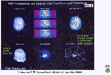
Figure DvC1-01 is from a Vital Images, Incorporated, VoxelGeo software advertisement (presently being developed and marketed by Paradigm Geophysical). It advertises their medical software (VoxelView), from which VoxelGeo was derived. Using MRI technology in this 1990 advertisement, a 3-D opaque human head is examined, virtually, showing the ability to control the opacity (transparency) of the individual Volume-PIXELs (aka, VOXELs) that make up a digital computerized volume. Here, “planting a seed” in a suspected tumor and performing “sub-volume detection” is demonstrated. This technique permits an interpreted 3-D opaque tumor to be viewed at any angle (and in animated motion), within a background of transparent VOXELs. This revolutionary 3D medical technology provided the impetus for transforming an unconventionally high-resolution, 2D seismic processing sequence into the patented D3DSP.
Like oil industry workers who deal with hidden targets in the visibly opaque earth, physicians and medical researchers cannot see inside the human body without visualization assistance. Their most common tools of choice are X-ray (Computerized Axial Tomography, or CAT) scans, induced-atomic-nuclei Magnetic-Resonance Imaging (MRI, using an injected paramagnetic dye), and diagnostic ultrasound measurements (medical "D3D seismic"), using both instantaneous snapshots and TV-monitored real-time movies. Positron Emission Tomography (PET scan), using an injected low-intensity-radioactive dye, is becoming another common tool for the medical arts. When a doctor wishes to investigate an object inside an opaque human body, they do not hire RN's to pick peaks and troughs on EKG's. Nor do they contract with processors to calculate MRI attribute volumes, or offer overrides to radiologists to make structure and amplitude maps from CAT scans. This display shows an example of a common use of appropriate medical imaging technology: an MRI intensity data volume of a patient's head. It is borrowed from the early Vital Images advertisement for a commercial volume-visualization computer program, called VoxelView (Vital Images, 1991). A "VOXEL" is defined as a "volumetric PIXEL", or volume "picture element". It is the elemental building block of any digital 3-D data set to be analyzed with volume visualization software (Vincent Argiro, 1990).
In this published commercial example, the shape of the opaque skulls (lower left) is examined by means of traditional planar slices to identify a suspected high-MRI-radiation-intensity anomaly (or tumor, upper-left and middle). The medical operator then chooses a "seed point", or starting VOXEL, on a slice through a potentially pathological subvolume (upper right). One by one, increasing intensity cutoff values are used and logical "detections" are performed, each time building a common-intensity-object, or detected subvolume, of all VOXELs that are contiguous to the starting VOXEL and satisfying the criterion that their intensity values are greater than the cutoff value. When the detection cutoff, or threshold, is too low (too relaxed), the detected object will include surrounding normal tissue. To arrive at a "final" 3-D object, the step-by-step, detect-and-evaluate procedure below might be followed (remember that these medical targets are generally higher intensity, whereas the D3DSP usually seeks lower D3D-impedance):
1. Start with the highest (most intense) cutoff value for the starting VOXEL, forming the minimum size Common-Intensity Object, or medical-CIO.
2. Decrease the cutoff value by one "step".
3. "Detect" all adjacent VOXELs whose amplitudes satisfy a greater-than-or-equal-to (GE) criterion, and electronically flag them, so that they are included in the growing surface layer of the expanding object. [Prior to each detection run, the entire volume is "reset", insuring that the intensity amplitude of every single VOXEL starts as an even integer. When any of the 26 surrounding and adjacent VOXELs is found to satisfy the GE criterion, it is said to be critically "detected", and the binary number "one" is added to the VOXEL's amplitude to make it an odd integer].
4. Increase the opacity of all detected VOXELs, forming a relatively opaque "common-intensity" object.
5. Evaluate the size, shape, location, and other quantitative attributes of the object.
6. Repeat steps 2-5 until the medical-CIO is too large to represent just the medical target (e.g., tumor, bone chip, etc.).
7. Reduce the cutoff value by one step, backing it off from its "overgrown size".
The resulting medical-CIO is usually the final diagnosed pathological tissue, as judged by an experienced physician or radiologist. It is the "intensity-cutoff interpretation" that will determine the significance (and accuracy) of all the following analyses and measurements, to be used for prescribing treatment or non-treatment. In the lower center and right corner of this Figure, the opaque object is the maximum size, anomalous, three-dimensional sub-volume that could be grown, and is therefore judged to be a "seed-filled" brain tumor, to be virtually measured and evaluated.
In the article seen under the VTV Home Page "Objects vs. Layers" button, the D3DSP "Objects vs. Layers" analogy is made to both the Wave-Particle Duality and to high-tech medical methods. In comparing these concepts, there is always the risk of seeming to overstate the importance of the D3DSP or seeming to trivialize the W-P Duality or the resulting medical tools. It is clear that the life-or-death importance of this final, volumetric, "intensity-cutoff interpretation" truly cannot be overstated (for the patient). It is of paramount importance to recognize the widely ignored (in the oil and gas industry) fact that digital data processing of complex 3D data volumes, always acts as a "pre-interpretation" of the medical- or geological-CIO's imbedded in a data volume. And this is much more critical in the seismic than in the medical theaters, because there are many more decisions made by a seismic processor (seeking the highest "layered-signal"-to-noise ratio), than by a medical data volume processor. The D3DSP contends that this is because medical workers know their targets are 3D objects and have no need to work so hard to create a "layered look".
|

